Knee
Management of an osteolytic lesion of the distal femur affecting the knee joint
This clinical case presents a 66-year-old patient suffering from mechanical pain in the left knee for several months. Clinical and imaging examinations reveal abnormalities that require thorough evaluation to determine the nature of the lesion.

Hopital Pierre Paul Riquet, Toulouse, France
Orthopaedic surgeon
Part one
Clinical presentation
- Male, 66 years old, hypertension, type 2 diabetes
- Mechanical pain in the left knee for 7–8 months prompting further investigations.
- Clinical examination: dry knee, 0/0/140°, localized tenderness on palpation of the medial femoral condyle
Imaging assessment
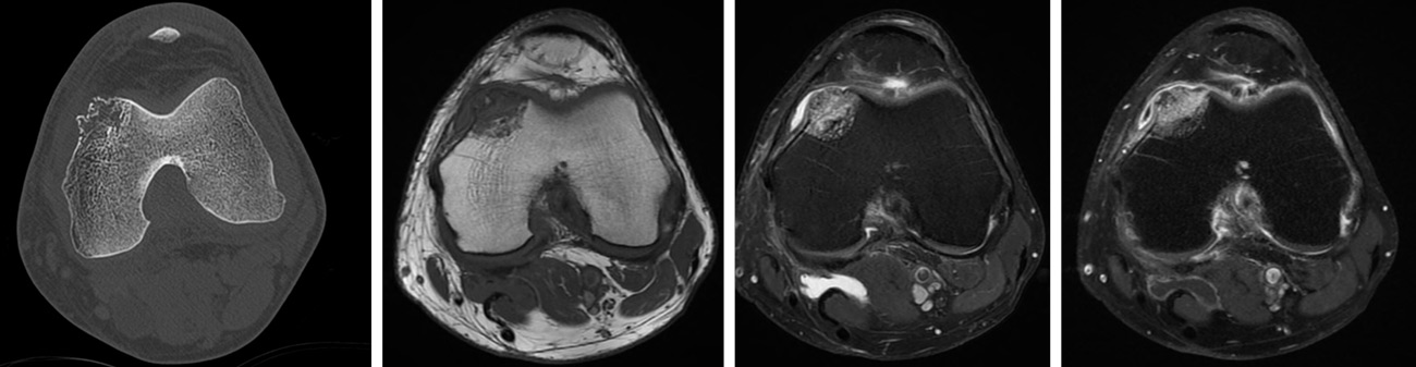
- CT-scan: Subchondral osteolytic lesion of the medial femoral condyle, with cortical breach.
- MRI: HypoT1, hyper T2, gado +
Part two
What is your diagnostic hypothesis?
- ✔️Cartilaginous tumor
How would you manage this condition?
- ✔️Oncologic resection
- ✔️Biopsy
- ✔️Multidisciplinary team meeting
Final strategy decision
A CT-guided Biopsy was done and an atypical cartilaginous tumor / low-grade chondrosarcoma was diagnosed.
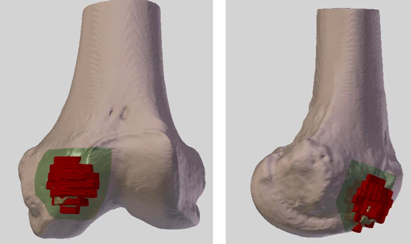
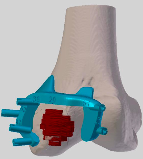
An oncologic resection using patient-specific cutting guides (3D-SIDE company) and reconstruction with allograft was performed.
The final pathology concluded to a grade 2 chondrosarcoma and a complete resection.
video
video
Comments
(No subject)
Nice job