In modern orthopedic surgery, over-reliance on magnetic resonance imaging (MRI) can obscure accurate diagnosis. In an interview with Matthieu Ollivier at PCO 2026, Sachin Tapasvi emphasizes why rigorous clinical examination remains the cornerstone of effective knee evaluation and patient-centered care.
Read more
Clinical diagnosis vs. imaging-first mindset
A comprehensive history and hands-on clinical assessment are essential before reviewing radiological data. While advanced imaging provides valuable confirmation, prioritizing MRI findings risks treating isolated radiological anomalies rather than the patient's actual symptomatic pathology.
Evaluating laxity patterns & surgical decision-making
- Laxity assessment: Physical tests reveal subtle laxity patterns, such as rotational instability or underlying hyperlaxity, that static imaging and laximeters cannot fully capture without side-to-side comparison.
- Surgical precision: Combining clinical findings with imaging ensures tailored surgical indications, preventing over-treatment and optimizing functional outcomes.
Both the Pune Knee Course and PCO reinforce this practical philosophy, focusing on real-life clinical scenarios, surgical indications, and hands-on technique over theoretical statistics.
Watch the full interview to understand why clinical examination remains essential before reviewing imaging.

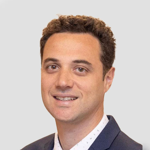
Matthieu Ollivier is currently a Professor of Orthopaedic Surgery at the Institute of Movement and Locomotion (Chairman: Pr Jean-Noel Argenson) at Aix-Marseille University Hospital, in Marseille, France. His clinical practice is dedicated to knee-related procedures, with a special focus on sports injuries (cartilage, meniscus, ligaments) and osteotomies. He is engaged in an academic field at Aix-Marseille University, teaching medical students, interns, orthopedic residents and fellows.