Knee
Femoral fracture unmasking dedifferentiated chondrosarcoma: A 52-year-old's diagnosis
What are the therapeutic options in case of spontaneous pathological diaphyseal fracture of the right femur leading to the diagnosis of a dedifferentiated chondrosarcoma in a 52-year-old man? How would you manage this clinical case?

Hopital Pierre Paul Riquet, Toulouse, France
Orthopaedic surgeon
Pierre Paul Riquet Hospital, Toulouse, France
Orthopaedic surgeon
Part one
Clinical presentation
- 52 year-old man
- Suffering bilateral leg pain for several months, diagnosed as a sciatic on the left side and as a truncated cruralgia on the right side.
- The compressive herniated disc L4/L5 was operated on the 30/01/2023
- A spontaneous, non-traumatic, fracture of the right femoral shaft occurred during the immediate post-operation manipulation (30/01/2023)
- The X-ray showed a pathological diaphyseal fracture of the right femur
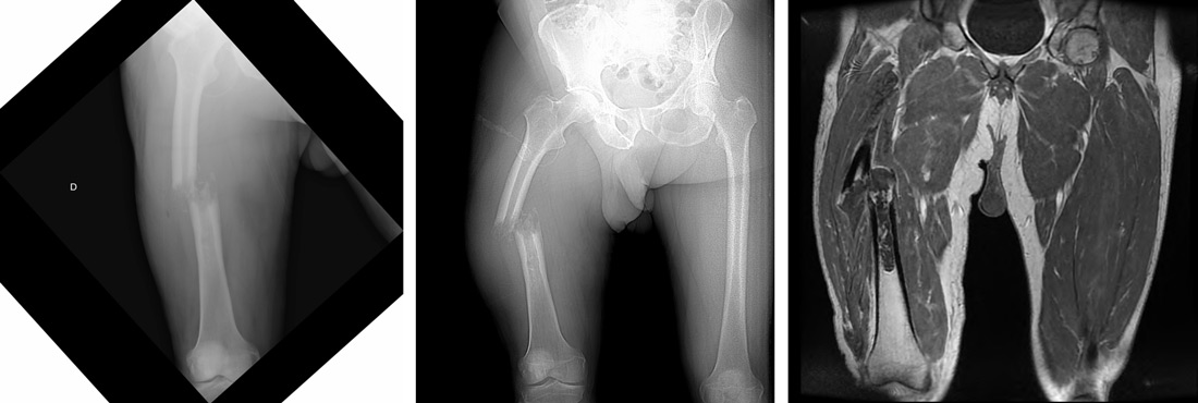
Further exams were done (CT, MRI, TEP-scan) and the case was discussed during the oncologic pluri-disciplinary meeting.
A biopsy of the lesion was done on the 10/02/2023, the analysis concluded to a dedifferentiated chondrosarcoma.
Part two
Quiz results
How would you manage this condition?
- ✔️Radiological biopsy, oncologic treatment and later removal and reconstructive tumoral surgery
Final strategy decision
- After anatomopathological results of the biopsy and discussion at the oncologic pluri-disciplinary meeting treatment plan was set up with neoadjuvant chemotherapy followed by surgery
- A trans-tibial traction of the leg was done, to try to relieve the fracture pain during the medical treatment time
- The follow-up scan at 4 months showed a very limited evolution of the tumor, without obvious regression and without consolidation of the fracture
- The operation was planned for 29/06/2023
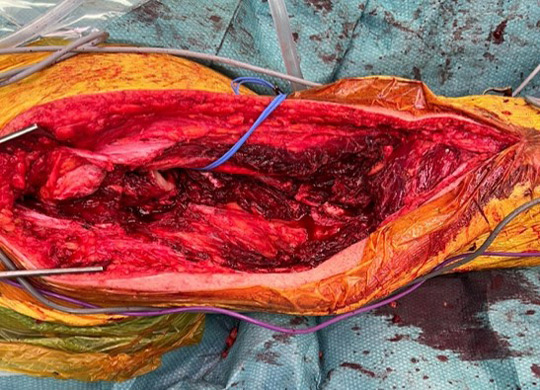
Surgery 06/2023
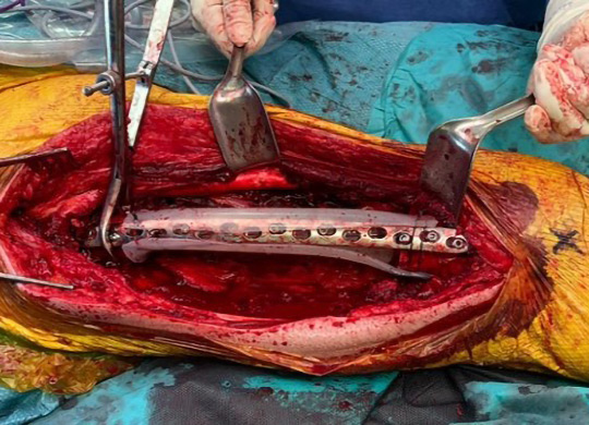
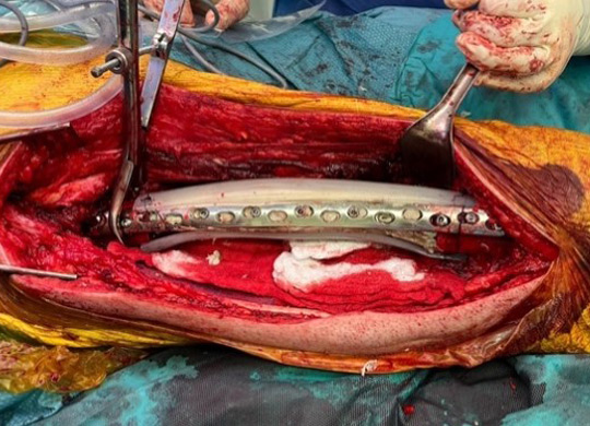
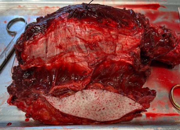
- A monobloc resection of the tumor was made with a femur proximal section at 7cm distal to the small trochanter and a distal section at 12cm of the interlining joint of the knee
- An important muscle portion of the quadriceps was removed
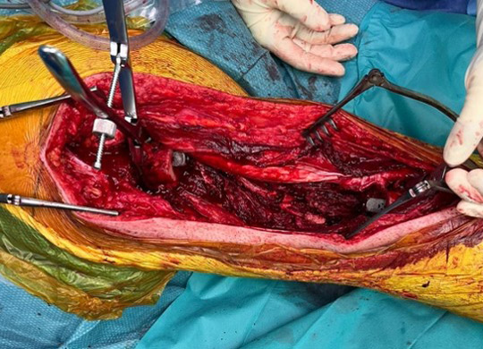
- The diaphysis was then reconstructed with a masquelet technique, divided into an endo-osteal and an extra-osteal fixation.
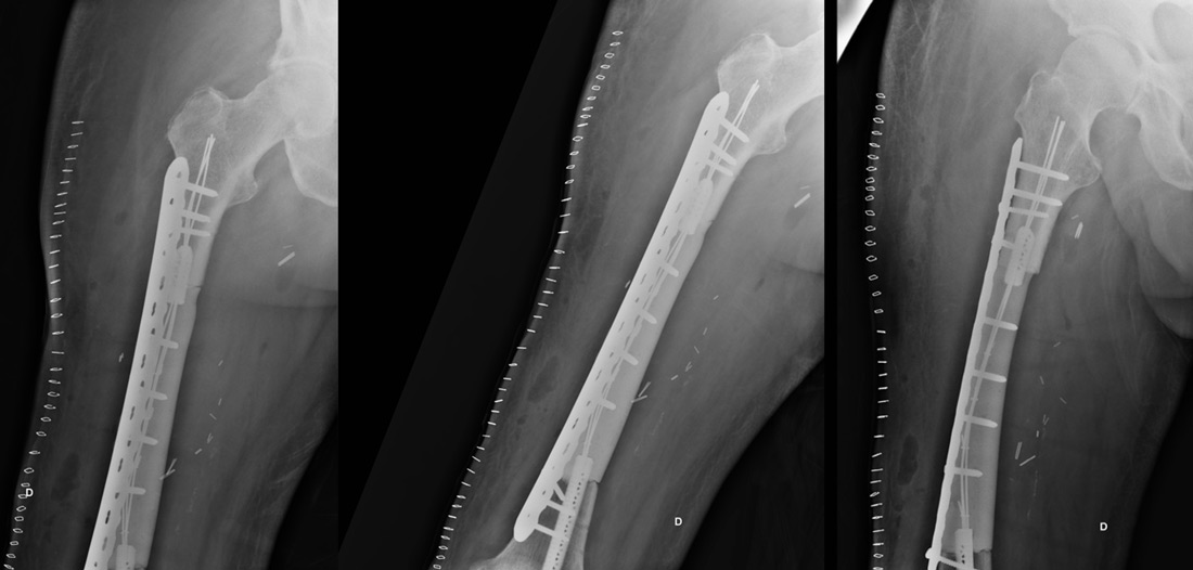
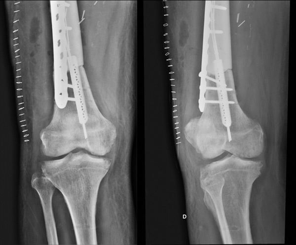
- For the internal fixation of the masquelet we used 2 Fuse-3D implants impacted in the proximal and distal remaining diaphysis of the femur and 3 spindles diameter 1,5, length 40cm were fixed inside and surrounded by wire cerclage
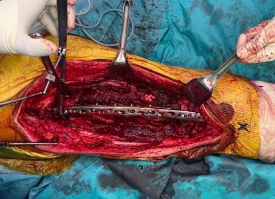
- For the external fixation of the masquelet we used a lateral femur plate fixed by 11 screws (7 fixed in the remaining bone, 4 fixed in the masquelet)
- Around this montage, cement was sunk
Per-op pictures
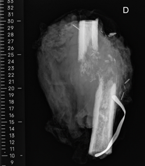
Post-op x-rays
Patient follow-up
- Anatomopathological results: R0, with bad chemotherapy response
- Pulmonary metastases were discovered and no reconstruction is scheduled for now
- Clinically: able to stand up and walk for a few steps with a walker
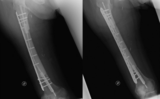
X-rays at 1 month 07/2023