A visual journey through orthopedic conditions. Discover the pathology in medical imaging, then in real life. A clear and focused perspective.
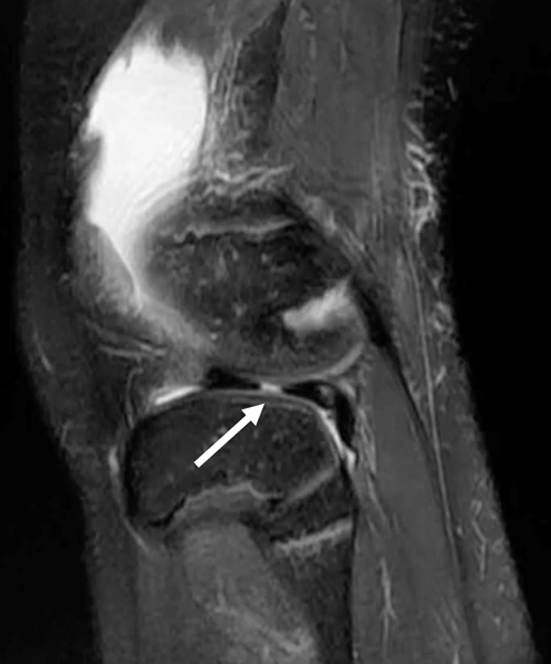
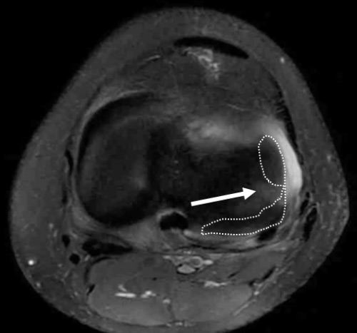
Accurate and timely diagnosis of lateral meniscus radial tears is crucial. These tears disrupt circumferential fibers, impairing meniscal function and joint stability. Often underdiagnosed on imaging, they require careful arthroscopic evaluation when suspected.


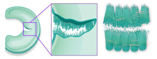
The proper management of lateral meniscus radial tears relies fundamentally on timely and accurate diagnosis. These lesions disrupt the integrity of the circumferential collagen fibers, which are essential for transmitting hoop stresses and maintaining meniscal load-bearing function.
They can lead to significant biomechanical impairment of the knee joint, increasing contact pressures and accelerating cartilage degeneration.
In the specific context of anterior cruciate ligament (ACL) reconstruction, lateral meniscus radial tears—especially oblique radial variants (LMORTs)—are relatively frequent and clinically relevant. If left untreated, they may contribute to persistent rotational instability and compromise graft protection. For this reason, identifying and repairing these lesions at the time of ACL reconstruction is critical to restore joint stability and improve long-term functional outcomes.
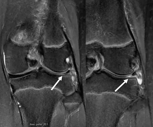
Radial tears, particularly complete or near-complete lesions in the midbody or posterior horn, often remain underdiagnosed on preoperative imaging, especially when subtle or associated with meniscal extrusion. Therefore, a high index of suspicion is essential, and MRI findings should be complemented by thorough arthroscopic evaluation of the lateral compartment to avoid missed diagnoses.
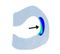
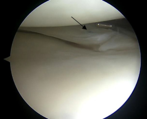
Anterior view of the lateral compartment of the knee with the diagnosis of a radial tear of the lateral meniscus
Anterior view of the lateral compartment of the knee with the diagnosis of a radial tear of the lateral meniscus
The all-inside repair with a side-to-side suture technique is an effective and minimally invasive option for treating radial tears of the lateral meniscus, particularly at the posterior horn or midbody. This approach allows restoration of the circumferential fiber continuity and meniscal function, providing good mechanical stability without the need for accessory incisions. When performed during ACL reconstruction, it contributes to joint stability and promotes healing.